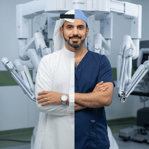
عبدالرحمن المحرج | Abdulrahman Almuhrij
- …
عبدالرحمن المحرج | Abdulrahman Almuhrij
- …
عبدالرحمن المحرج | Abdulrahman Almuhrij
نصائح طبية
Medical Advice
Kidney and Ureter Stones
Kidney stones are one of the most common urological conditions, affecting up to 1 in 10 people during their lifetime. As a urology consultant subspecialized in endourology, I treat kidney and ureter stones regularly using minimally invasive techniques — and most cases are entirely preventable with the right habits.
**What causes kidney stones?**
Stones form when minerals and salts in urine — most commonly calcium oxalate — crystallize and clump together. Risk factors include dehydration, high-salt diets, excess animal protein, family history, and certain medical conditions like obesity or gout.
**Common symptoms**
- Severe pain in the side or back, below the ribs
- Pain that radiates to the lower abdomen and groin
- Blood in the urine
- Nausea and vomiting
- Frequent, painful urination
If you experience sudden, severe flank pain, seek medical attention promptly — this is one of the most painful conditions in medicine, and early treatment prevents complications like kidney damage or infection.
**Prevention: what actually works**
Drink plenty of fluid: 2-2.5 Liters/day. Aim to make urine color clear or cloudlessLimit foods with high oxalate content (example: dark chocolate, peanuts, rhubarb, spinach, beets, Swiss chard, berries, such as strawberries, raspberries, and blackberries and sweet potatoes). This helps calcium oxalate stone patients
Eat enough dietary calcium (Normal level). The recommended calcium intake to prevent calcium stones is 1000-1200 mg per day. This helps calcium oxalate stone patients
For uric acid stone formers, eat a Low to moderate amount of protein
Low salt intake. Extra sodium causes you to lose more calcium in your urine. There are many sources of "hidden" sodium such as canned or commercially processed foods as well as restaurant-prepared and fast foods. This helps calcium oxalate stone patients
Avoid high doses of vitamin C supplements. 60mg/day of vitamin C. Excess amounts of 1000mg/day or more may produce more oxalate in the body
General health: overweight people and sedentary occupations or lifestyles
**Modern treatment options**
Most stones under 5mm pass on their own with hydration and pain control. Larger or symptomatic stones may need intervention:
- **Shockwave lithotripsy (ESWL)** — non-invasive sound waves break the stone, lower success but less complications rate
- **Ureteroscopy with laser** — a thin scope removes or breaks up the stone through natural passages, no incisions. Most stones treated with this approach.
- **Percutaneous nephrolithotomy (PCNL)** — for large or complex stones, via a small keyhole access
Modern endourology means most patients go home the same day or after one night, with minimal recovery time.
**When to see a urologist**
If you've had a kidney stone before, you have roughly a 50% chance of another within 5-10 years without preventive changes. A metabolic evaluation can identify your specific risk factors and guide targeted prevention.
حصوات الكلى والحالب
تُعد حصوات الكلى من أكثر الحالات البولية شيوعًا، حيث تصيب ما يصل إلى واحد من
كل عشرة أشخاص خلال حياتهم. بصفتي استشاري جراحة مسالك بولية متخصص دقيق في المناظير البولية، أعالج حصوات الكلى والحالب بانتظام باستخدام تقنيات طفيفة التوغل — ومعظم الحالات يمكن الوقاية منها تمامًا باتباع عادات صحية صحيحة.
**ما الذي يسبب حصوات الكلى؟**
تتكون الحصوات عندما تتبلور الأملاح والمعادن الموجودة في البول — وأكثرها شيوعًا أكسالات الكالسيوم — وتتجمع معًا. تشمل عوامل الخطر الجفاف، والأنظمة الغذائية عالية الملح، والإفراط في البروتين الحيواني، والتاريخ العائلي، وبعض الحالات الصحية مثل السمنة والنقرس.
**الأعراض الشائعة**
- ألم شديد في الجانب أو الظهر أسفل الأضلاع
- ألم ينتشر إلى أسفل البطن والفخذ
- دم في البول
- غثيان وقيء
- تبول متكرر ومؤلم
إذا شعرت بألم مفاجئ وشديد في الخاصرة، فاطلب الرعاية الطبية فورًا — فهذه من أكثر الحالات إيلامًا في الطب، والعلاج المبكر يمنع المضاعفات مثل تلف الكلى أو العدوى.
**الوقاية: ما الذي يجدي فعلًا**
شرب كمية كافية من السوائل: 2-2.5 لتر خلال اليوم. الهدف هو تقليل تركيز الأملاح في البول وجعل البول لون أصفر فاتح أو شبة شفاف
قم بتقليل الأطعمة التي تحتوي على أوكساليت (أمثلة: الشوكولاته الداكنة، بعض أنوع المكسرات، السبانخ، التوت والفراولة والبطاطا الحلوة). خاصة لمرضى حصوات الكالسيوم أوكساليت
قم باستهلاك كمية صحية وكافية من الكالسيوم بشكل يومي، لا تقم بتقليل الكمية أو زيادتها عن الحاجة الطبيعية. استهدف مستوى 1000-1200 مل جرام بشكل يومي من أصناف الأغذية أو المكملات الصحية. خاصة لمرضى حصوات الكالسيوم أوكساليت
لمرضى حصوات حمض اليورك، قم باستهلاك كمية بسيطة إلى متوسطة من البروتين من اللحوم بشكل خاص، كثرتها تزيد من نسبة تكون الحصوات
قلل من استهلاك الملح بشكل يومي. كثرة ملح الصوديوم يزيد كثرة الكالسيوم في البول مما يزيد نسبة احتمال تكون الحصوات. الأملاح تكون بشكل عالي في الوجبات الجاهزة والمعلبة والمطاعم. خاصة لمرضى حصوات الكالسيوم أوكساليت
قم بتجنب الجرعات العالية من فيتامين ج (C). الهدف هو حصول الجسم على 60 مل جرام بشكل يومي. الكميات العالية في الجسم قد تجعل مستوى الأوكسالات أعلى في البول مما يزيد نسبة احتمال تكون الحصوات
اهتم بصحتك بشكل عام: زيادة الوزن والحياة الغير نشطة قد تزيد من نسبة تكون الحصوات
**خيارات العلاج الحديثة**
معظم الحصوات التي يقل حجمها عن 5 ملم تخرج تلقائيًا مع شرب الماء ومسكنات الألم. أما الحصوات الأكبر أو المصحوبة بأعراض فقد تحتاج إلى تدخل.
- **تفتيت الحصوات بالموجات الصادمة (ESWL)** — تقنية غير جراحية تستخدم الموجات الصوتية لتفتيت الحصوة. أقلهم نجاحا في علاج الحصوات لكن أقلهم نسبة مضاعفات.
- **المنظار الحالبي بالليزر** — منظار رفيع يزيل الحصوة أو يفتتها عبر المسالك الطبيعية دون شقوق جراحية. هذا الخيار الأولي لعلاج أغلب حالات الحصوات.
- **استخراج حصوات الكلى عبر الجلد (PCNL)** — للحصوات الكبيرة أو المعقدة، عبر فتحة صغيرة
بفضل تقنيات المناظير البولية الحديثة، يعود معظم المرضى إلى منازلهم في نفس اليوم أو بعد ليلة واحدة، مع فترة تعافٍ قصيرة.
**متى تزور طبيب المسالك البولية**
إذا سبق أن أصبت بحصوة كلى، فاحتمال إصابتك بأخرى خلال 5-10 سنوات يبلغ نحو 50% دون اتخاذ إجراءات وقائية. يمكن للتقييم الأيضي تحديد عوامل الخطر الخاصة بك وتوجيه خطة وقاية مناسبة لحالتك.
Why We Choose Robotic Surgery
Robotic surgery has transformed urology over the past decade. As a urologist trained in robotic and laparoscopic techniques, I use these approaches for a wide range of conditions — from kidney tumors to prostate cancer — to reduce pain, scarring, and recovery time compared to traditional open surgery.
Robotic surgery not only for cancer, but also for benign condition like narrowing of the ureter or issue with bladder and prostate.
Robotic surgery is a great option for pediatric age group.
**What's the difference?**
- **Laparoscopic surgery** uses small incisions (usually under 1cm) and long, thin instruments guided by a camera. The surgeon operates directly.
- **Robotic-assisted surgery** uses small incisions, but the surgeon controls robotic arms from a console, offering 3D vision and greater precision, especially in tight anatomical spaces like the pelvis.
Both are true minimally invasive techniques — the "robot" doesn't operate independently; it's a precision tool controlled entirely by the surgeon.
**Common procedures using these techniques**
- **Radical/partial nephrectomy** — kidney tumor removal, often preserving healthy kidney tissue
- **Radical prostatectomy** — prostate cancer surgery with better nerve-sparing precision
- **Pyeloplasty** — repairing blockages between kidney and ureter
- **Adrenalectomy** — adrenal gland tumor removal
**Ureter reconstruction** — treating narrowed Ureter by reconstruction
**Benefits compared to open surgery**
- Smaller incisions, minimal scarring
- Less blood loss
- Significantly less post-operative pain
- Shorter hospital stay (often 1-2 days vs. 1-2weeks)
- Faster return to normal activity and work
**Is it right for everyone?**
Not every case is suited to a minimally invasive approach — tumor size, location, and a patient's overall health all factor in. A proper evaluation with imaging determines the safest and most effective option for your specific situation.
**What to expect**
Most patients walk within a day of surgery and go home within 1-2 days for standard procedures. Full recovery to normal activity typically takes 2-4 weeks, far faster than the 8-10 weeks common with open surgery.
لماذا نختار جراحة الروبوت / الرجل الآلي
غيّرت الجراحة الروبوتية مجال المسالك البولية جذريًا خلال العقد الماضي. بصفتي طبيب متخصص بالروبوت ومدرب للجيل الجديد من الاطباء على تقنيات الجراحة الروبوتية والمنظارية، أستخدم هذه الأساليب لعلاج مجموعة واسعة من الحالات — من أورام الكلى إلى سرطان البروستاتا — لتقليل الألم والندوب وفترة التعافي مقارنة بالجراحة المفتوحة التقليدية.
الروبوت ليس فقط لجراحة الأورام، يتم استخدامه لعلاج أي من المشاكل الصحية الحميدة مثل مشاكل الحالب البولي والمثانة والبروستاتا.
عمليات الروبوت هي خيار جيد وآمن للأطفال.
**ما الفرق؟**
- **الجراحة بالمنظار** تستخدم شقوقًا صغيرة (عادة أقل من 1 سم) وأدوات طويلة ورفيعة موجهة بكاميرا، ويتحكم الجراح بها مباشرة.
- **الجراحة بمساعدة الروبوت** تستخدم أيضًا شقوقًا صغيرة، لكن الجراح يتحكم في الأذرع الروبوتية من طاولة تحكم، مما يوفر رؤية ثلاثية الأبعاد ودقة أعلى، خاصة في المناطق التشريحية الضيقة كالحوض.
كلتا التقنيتين جراحة طفيفة التوغل حقيقية — فـ"الروبوت" لا يعمل بشكل مستقل، بل هو أداة دقيقة يتحكم بها الجراح بالكامل.
**الإجراءات الشائعة التي تستخدم هذه التقنيات**
- **استئصال الكلى الجزئي أو الكامل** — إزالة أورام الكلى مع الحفاظ على الأنسجة السليمة قدر الإمكان
- **استئصال البروستاتا الجذري** — جراحة سرطان البروستاتا بدقة أعلى في الحفاظ على الأعصاب
- **رأب الحويضة** — إصلاح انسدادات بين الكلى والحالب
- **استئصال الغدة الكظرية** — إزالة أورام الغدة الكظرية
- **إصلاح مجرى الحالب** — معالجة تضيق الحالب
**الفوائد مقارنة بالجراحة المفتوحة**
- شقوق أصغر وندوب أقل
- فقدان دم أقل
- ألم أقل بشكل ملحوظ بعد الجراحة
- إقامة أقصر في المستشفى (غالبًا يوم إلى يومين مقابل أسبوع إلى أسبوعين)
- عودة أسرع للنشاط الطبيعي والعمل
**هل هي مناسبة للجميع؟**
ليست كل الحالات مناسبة للأسلوب طفيف التوغل — فحجم الورم وموقعه والحالة الصحية العامة للمريض كلها عوامل مؤثرة. يحدد التقييم الدقيق مع الأشعة الخيار الأكثر أمانًا وفعالية لحالتك.
**ماذا تتوقع**
يمشي معظم المرضى خلال يوم من الجراحة ويعودون لمنازلهم خلال يوم إلى يومين للإجراءات المعتادة. يستغرق التعافي الكامل للنشاط الطبيعي عادة 2-4 أسابيع، أسرع بكثير من 8-10 أسابيع المعتادة مع الجراحة المفتوحة.
Prostate Biopsy
Transperineal Prostate Biopsy: Why It's Now the Preferred Approach
Prostate biopsy is the standard way to confirm a prostate cancer diagnosis when imaging or PSA levels raise suspicion. For decades, this was almost always done transrectally (through the rectum). Today, transperineal biopsy — performed through the skin between the scrotum and anus — has emerged as the preferred technique in most major urology guidelines, and it's the approach I now recommend for my patients whenever appropriate.
**What's the difference between the two techniques?**
- **Transrectal biopsy (TRUS)**: the traditional approach. An ultrasound probe is placed in the rectum, and biopsy needles pass through the rectal wall to sample the prostate.
- **Transperineal biopsy**: the needle instead passes through the perineal skin (between the scrotum and anus) — it never touches the rectum at all.
**Why transperineal has become preferred**
- **Lower infection risk** — this is the main driver behind the shift. Passing needles through the rectum carries bacteria into the prostate and bloodstream, which can cause infection or, in rare cases, life-threatening sepsis. The transperineal route avoids the rectal wall entirely, substantially lowering this risk.
- **Comparable or better cancer detection**research comparing the two approaches consistently shows similar or improved detection of clinically significant prostate cancer with the transperineal technique, particularly for tumors in the front (anterior) part of the prostate, which the transrectal route can under-sample.
- **Reduced need for antibiotics** — because infection risk is lower, some transperineal biopsies can be performed with reduced or even no antibiotic prophylaxis, which also helps limit antibiotic resistance.
- **Guideline recommendation** — European urology guidelines now strongly recommend transperineal biopsy as the preferred, guideline-endorsed approach. Some other major guidelines still consider both approaches acceptable, but the trend across the field is clearly moving toward transperineal.
**How the procedure is done**
Modern transperineal biopsy is typically performed under local anesthesia in an outpatient setting — not necessarily requiring general anesthesia or a hospital stay, using a small grid template or freehand technique guided by ultrasound (often combined with MRI fusion for precise targeting of suspicious areas). Most patients tolerate it well, describe it as no more uncomfortable than a transrectal biopsy, and go home the same day.
**What to expect afterward**
Mild soreness in the perineal area for a few days, and occasionally minor blood in the urine or semen — this is normal and resolves on its own. Recovery is typically quick, and most patients return to normal activity within a day or two.
**The bottom line**
If you're facing a recommendation for a prostate biopsy, ask your urologist about the transperineal approach — it offers a meaningfully lower infection risk with equal or better diagnostic accuracy, which is why it's increasingly becoming the standard of care.
This technique is available and I performed more than 350 cases without infection complications and with better diagnosis, 98% of these cases performed under local anaesthsia.
خزعة البروستاتا
خزعة البروستاتا عبر العجان: النهج المفضل حديثًا
منطقة العجان هي منطقة الجلد الخارجية التي تقع بين كيس الصفان (الخصيتين) و فتحة التبرز.
خزعة البروستاتا هي الطريقة المعتمدة لتأكيد تشخيص سرطان البروستاتا عندما تثير الأشعة أو مستوى مستضد البروستاتا النوعي (PSA) الشك. لعقود طويلة، كانت تُجرى غالبًا عبر المستقيم. اليوم، أصبحت الخزعة عبر العجان — التي تتم عبر الجلد بين كيس الصفن والشرج — النهج المفضل في معظم الإرشادات الطبية الكبرى لجراحة المسالك البولية، وهي الطريقة التي أوصي بها الآن لمرضاي كلما كان ذلك مناسبًا.
**ما الفرق بين الطريقتين؟**
- **الخزعة عبر المستقيم (TRUS)**: الطريقة التقليدية. يوضع مسبار السونار داخل المستقيم، وتمر إبر الخزعة عبر جدار المستقيم لأخذ عينات من البروستاتا.
- **الخزعة عبر العجان**: تمر الإبرة بدلًا من ذلك عبر جلد العجان (المنطقة بين كيس الصفن والشرج) — ولا تلامس المستقيم إطلاقًا.
**لماذا أصبحت الخزعة عبر العجان هي المفضلة**
- **خطر عدوى أقل** — وهذا هو الدافع الرئيسي وراء هذا التحول. مرور الإبر عبر المستقيم ينقل البكتيريا إلى البروستاتا ومجرى الدم، مما قد يسبب التهابًا أو في حالات نادرة تسممًا دمويًا خطيرًا. تتجنب الطريقة العجانية جدار المستقيم تمامًا، مما يقلل هذا الخطر بشكل كبير.
- **دقة تشخيصية مماثلة أو أفضل** — تُظهر الأبحاث التي قارنت بين الطريقتين باستمرار كشفًا مماثلًا أو أفضل لسرطان البروستاتا ذي الأهمية السريرية بالطريقة العجانية، خاصة للأورام في الجزء الأمامي من البروستاتا، والتي قد لا تُؤخذ منها عينات كافية بالطريقة عبر المستقيم.
- **حاجة أقل للمضادات الحيوية** — نظرًا لانخفاض خطر العدوى، يمكن إجراء بعض الخزعات العجانية بمضادات حيوية وقائية أقل أو حتى دونها، مما يساعد أيضًا في الحد من مقاومة المضادات الحيوية.
- **توصيات الإرشادات الطبية** — توصي الإرشادات الأوروبية لطب المسالك البولية الآن بقوة بالخزعة عبر العجان كالنهج المفضل والمعتمد. لا تزال بعض الإرشادات الكبرى الأخرى تعتبر الطريقتين مقبولتين، لكن الاتجاه العام في هذا المجال يتحرك بوضوح نحو الخزعة العجانية.
**كيف يتم الإجراء**
تُجرى الخزعة العجانية الحديثة عادة تحت تخدير موضعي في العيادة الخارجية — دون الحاجة بالضرورة إلى تخدير عام أو إقامة في المستشفى، باستخدام قالب شبكي صغير أو تقنية حرة موجهة بالسونار (وغالبًا ما تُدمج مع دمج صور الرنين المغناطيسي لاستهداف دقيق للمناطق المشتبه بها). يتحمل معظم المرضى الإجراء بشكل جيد، ويصفونه بأنه لا يزيد إزعاجًا عن الخزعة عبر المستقيم، ويعودون إلى منازلهم في نفس اليوم.
**ماذا تتوقع بعد الإجراء**
ألم خفيف في منطقة العجان لبضعة أيام، وأحيانًا دم بسيط في البول أو السائل المنوي — وهذا أمر طبيعي يزول من تلقاء نفسه. يكون التعافي سريعًا عادة، ويعود معظم المرضى لنشاطهم الطبيعي خلال يوم أو يومين.
**خلاصة القول**
إذا أوصى طبيبك بإجراء خزعة للبروستاتا، اسأله عن الخزعة عبر العجان — فهي توفر خطر عدوى أقل بشكل ملحوظ مع دقة تشخيصية مماثلة أو أفضل، ولهذا السبب أصبحت تدريجيًا المعيار المتبع في الرعاية الطبية.
التقنية متوفرة وفمت بعمل فوق 350 خزعة من هذه التقنية من دون مضاعفات التهاب وتشخيص بدقة أفضل، 98٪ من الحالات تم عملها بواسطة بنج موضعي.
Benign Prostate Enlargment
Prostate gland is only found in males. It is expected to get enlarged with age. It is part of the male urethra (water pipe) and with its enlargment, it can cause narrowing in the urethra and make voiding issues
A non-cancerous enlargement of the prostate — affects most men to some degree after age 50. It's extremely common, highly treatable, and not something to be embarrassed about discussing.
**Common symptoms**
- Frequent urination, especially at night (nocturia)
- Weak or interrupted urine stream
- Difficulty starting urination
- Feeling the bladder isn't fully empty
- Sudden, urgent need to urinate
These symptoms build gradually, which is why many men live with them for years before seeking help — often unnecessarily, since effective treatments exist at every stage.
**Diagnosis**
A proper evaluation includes a symptom questionnaire, urine flow test, PSA blood test, and sometimes an uroflowmetry test and ultrasound to measure prostate size and check for residual urine. This helps rule out other causes, including prostate cancer, and tailor treatment to your specific case.
Extra special tests may get requested like urodynamics and check cystosocpy.
**Treatment options, from conservative to advanced**
- **Lifestyle changes** — reducing evening fluids, limiting caffeine/alcohol
- **Medications** — alpha-blockers relax the prostate muscle for quick symptom relief; 5-alpha reductase inhibitors shrink the prostate over months
- Minimally invasive procedures:
Laser prostate surgery (HoLEP/GreenLight): remove excess tissue with less bleeding and faster recovery than traditional surgery
Water vaporization (REZUM): Effective treatment option for most cases
TransUrethral Resection of the Prostate (TURP): effective for most cases
Robotic simple prostatectomy: great option for large or complicted cases
Most men do very well with medication alone initially. Procedures are considered when symptoms significantly affect quality of life or medication isn't enough.
**When to see a urologist**
Don't wait until symptoms become severe. Untreated enlarged prostate can, in some cases, lead to urinary retention, bladder or kidney damage, or recurrent infections. An early evaluation is quick and can save years of unnecessary discomfort.
تضخم البروستاتا الحميد
يُعد تضخم البروستاتا الحميد — وهو تضخم غير سرطاني في غدة البروستاتا — حالة شائعة جدًا تصيب معظم الرجال بدرجة ما بعد سن الخمسين. هي حالة شائعة وقابلة للعلاج بشكل كبير، وليست أمرًا يستدعي الحرج من الحديث عنه.
غدة البروستاتا موجودة لدى الذكور فقط. من المتوقع تضخمها مع التقدم في العمر. البروستاتا جزء من مجرى التبول وتضخمها قد يؤدي إلى تضيق مجرى البول مما قد يؤدي إلى مشاكل في التبول
**الأعراض الشائعة**
- التبول المتكرر، خاصة ليلًا
- ضعف أو تقطع تيار البول
- صعوبة في بدء التبول
- الشعور بعدم إفراغ المثانة بالكامل
- الحاجة المفاجئة والملحة للتبول
تتطور هذه الأعراض تدريجيًا، ولهذا يعيش كثير من الرجال معها لسنوات قبل طلب المساعدة — وغالبًا دون داعٍ، لأن هناك علاجات فعالة في كل مرحلة.
**التشخيص**
يشمل التقييم السليم استبيان الأعراض، واختبار تدفق البول، وفحص دم لإنزيم البروستاتا (PSA)، واختبار دفع البول وأحيانًا الموجات فوق الصوتية لقياس حجم البروستاتا والتحقق من البول المتبقي. يساعد ذلك في استبعاد أسباب أخرى، بما فيها سرطان البروستاتا، وتحديد العلاج الأنسب لحالتك.
قد يتم طلب فحوصات إضافية خاصة مثل اختبار دينامكية التبول وعمل منظار تشخيصي لمجري لببول والبروستاتا والمثانة البولية.
**خيارات العلاج، من الأبسط إلى الأكثر تقدمًا**
- **تعديل نمط الحياة** — تقليل السوائل مساءً، والحد من الكافيين والكحول
- **الأدوية** — علاجات تستخدم لتُرخي عضلة البروستاتا لتخفيف سريع للأعراض؛ وعلاجات تُقلّص حجم البروستاتا خلال أشهر
- الإجراءات بسيطة التدخل:
- **جراحة الليزر للبروستاتا (HoLEP/GreenLight)**— التي تزيل الأنسجة الزائدة بنزيف أقل وتعافٍ أسرع من الجراحة التقليدية
- **التبخير بالماء (REZUM)** — خيار يناسب أغلب الحالات ويتميز بالتعافي السريع
- **جراحة تجريف/كحت البروستاتا بالمنظار (TURP)** — خيار فعال في أغلب الحالات
- **إزالة البروستاتا بالروبوت الجراحي (Robotic simple prostatectomy)** — خيار للحالات ذات الحجم الكبير والمعقد
يستجيب معظم الرجال جيدًا للأدوية وحدها في البداية. تُلجأ الإجراءات الجراحية عند تأثر جودة الحياة بشكل كبير أو عدم كفاية الأدوية.
**متى تزور طبيب المسالك البولية**
لا تنتظر حتى تصبح الأعراض شديدة. في بعض الحالات، قد يؤدي تضخم البروستاتا غير المعالج إلى احتباس بولي أو تلف في المثانة أو الكلى أو التهابات متكررة. التقييم المبكر سريع وقد يوفر عليك سنوات من الانزعاج غير الضروري.
Urology Cancer
Urologic cancers — of the prostate, kidney, ureter, adrenal, testis and bladder — are among the most common cancers, and also among the most treatable when caught early. Awareness and timely screening make the biggest difference in outcomes.
**Prostate cancer**
The most common cancer in men. Often has no symptoms in early stages, which is why screening matters. Risk increases with age (especially after 50) and family history. Screening involves a PSA blood test and, when needed, further evaluation. Treatment ranges from active surveillance for low-risk cases to robotic surgery or radiation for higher-risk disease — many men are cured with modern treatment.
**Kidney cancer**
Often discovered incidentally on imaging done for other reasons, since early kidney cancer typically causes no symptoms. Warning signs when present include blood in the urine, persistent flank pain, or an unexplained lump. Small kidney tumors are increasingly treated with partial nephrectomy — removing just the tumor and preserving healthy kidney function — using robotic or laparoscopic techniques.
**Bladder cancer**
The most common warning sign is blood in the urine — even a single episode, painless or not, should always be evaluated. Smoking is the single biggest risk factor. Diagnosis typically involves cystoscopy (a camera examination of the bladder). Early-stage bladder cancer has excellent treatment outcomes.
**Ureter - Transitional cell cancer**
Arises in the lining of the renal pelvis or ureter, sharing common traits with bladder cancer. The primary warning sign is blood in the urine, often accompanied by persistent flank pain or discomfort. Smoking and long-term chemical exposure are major risk factors. Diagnosis relies on imaging and ureteroscopy, with treatment typically involving surgical removal of the tumor or affected ureter while preserving kidney function whenever possible.
**Adrenal Cancer**
A rare cancer that develops in the adrenal glands sitting atop the kidneys. It is frequently discovered through hormonal imbalances—causing symptoms like sudden high blood pressure, unexpected weight gain, or muscle weakness—or incidentally on abdominal imaging. Surgical removal of the adrenal gland (adrenalclectomy) is the primary treatment, often combined with targeted therapies or medications to manage hormone levels.
**Testicular cancer**
The most common cancer in young men aged 15 to 35, highly treatable and often curable even when detected at later stages. Key signs include a painless lump, swelling, or a feeling of heaviness in the scrotum. Diagnosis involves a physical exam, ultrasound, and specific blood markers. Treatment usually starts with surgical removal of the affected testicle, followed by surveillance, chemotherapy, or radiation depending on the tumor type.
**The common thread: don't ignore these signs**
- Blood in urine (even once)
- Unexplained flank or lower back pain
- Changes in urination that persist beyond a few weeks
- A family history of urologic cancer
None of these symptoms automatically mean cancer — most don't — but they all warrant a proper evaluation rather than assumption or delay.
**Screening recommendations**
Men over 50 (or over 45 with family history or African ancestry) should discuss PSA screening with a urologist. Anyone with persistent urinary symptoms or visible blood in urine, regardless of age, should be evaluated promptly.
أورام المسالك السرطانية
تعد سرطانات المسالك البولية — البروستاتا والكلى والمثانة والغدة الكضرية والحالب والخصيتين — من أكثر أنواع السرطان شيوعًا، وأيضًا من أكثرها قابلية للعلاج عند اكتشافها مبكرًا. الوعي والفحص المبكر في الوقت المناسب يُحدثان الفرق الأكبر في النتائج.
**سرطان البروستاتا**
أكثر أنواع السرطان شيوعًا لدى الرجال. غالبًا لا يسبب أعراضًا في مراحله المبكرة، ولهذا يُعد الفحص المبكر مهمًا. يزداد الخطر مع التقدم في العمر (خاصة بعد سن الخمسين) والتاريخ العائلي. يشمل الفحص تحليل دم لمستضد البروستاتا النوعي (PSA)، وعند الحاجة، مزيدًا من التقييم. تتراوح خيارات العلاج من المراقبة النشطة للحالات منخفضة الخطورة إلى الجراحة الروبوتية أو العلاج الإشعاعي للحالات الأعلى خطورة — ويُشفى كثير من الرجال بالعلاج الحديث.
**سرطان الكلى**
غالبًا ما يُكتشف صدفة أثناء أشعة تُجرى لأسباب أخرى، لأن سرطان الكلى المبكر عادة لا يسبب أعراضًا. من العلامات التحذيرية عند ظهورها: دم في البول، ألم مستمر في الخاصرة، أو كتلة غير مفسرة. تُعالج أورام الكلى الصغيرة بشكل متزايد عبر استئصال جزئي للكلى — إزالة الورم فقط مع الحفاظ على وظيفة الكلى السليمة — باستخدام تقنيات روبوتية أو منظارية.
**سرطان المثانة**
أكثر العلامات التحذيرية شيوعًا هي الدم في البول — حتى لو حدث مرة واحدة وبدون ألم، يجب دائمًا تقييمه. التدخين هو أكبر عامل خطر منفرد. يشمل التشخيص عادة تنظير المثانة (فحص بالكاميرا). لسرطان المثانة في مراحله المبكرة نتائج علاجية ممتازة.
**سرطان الحالب وحوض الحالب**
ينشأ في البطانة الداخلية لحوض الكلية أو الحالب، ويشارك سرطان المثانة في العديد من الخصائص. العلامة التحذيرية الرئيسية هي وجود دم في البول، وغالباً ما يكون مصحوباً بألم مستمر في الخصر أو شعور بعدم الراحة. يُعد التدخين والتعرض الكيميائي طويل الأمد من أكبر عوامل الخطر. يعتمد التشخيص على التصوير الطبي وتنظير الحالب، وعادة ما يتضمن العلاج الاستئصال الجراحي للورم أو الحالب المصاب مع الحفاظ على وظيفة الكلى قدر الإمكان.
**سرطان الغدة الكظرية**
سرطان نادر يتطور في الغدد الكظرية الموجودة فوق الكليتين. غالباً ما يتم اكتشافه من خلال الاختلالات الهرمونية — مما يسبب أعراضاً مثل الارتفاع المفاجئ في ضغط الدم، أو زيادة الوزن غير المبررة، أو ضعف العضلات — أو بالصدفة عبر تصوير البطن. يُعد الاستئصال الجراحي للغدة الكظرية العلاج الأساسي، وغالباً ما يُدمج مع العلاجات الموجهة أو الأدوية للسيطرة على مستويات الهرمونات.
سرطان الخصية
أكثر أنواع السرطان شيوعاً بين الشباب الذين تتراوح أعمارهم بين 15 و35 عاماً، وهو قابل للعلاج بدرجة عالية وغالباً ما يُشفى تماماً حتى عند اكتشافه في مراحل متأخرة. تشمل العلامات الرئيسية ظهور كتلة غير مؤلمة، أو تورم، أو شعور بالثقل في كيس الصفن. يتضمن التشخيص الفحص السريري، والتصوير بالموجات الصوتية (السونار)، وفحوصات دلالات الأورام في الدم. يبدأ العلاج عادة بالاستئصال الجراحي للخصية المصابة، يليه المراقبة أو العلاج الكيميائي أو الإشعاعي اعتماداً على نوع الورم.
**القاسم المشترك: لا تتجاهل هذه العلامات**
- دم في البول (حتى لو مرة واحدة)
- ألم غير مفسر في الخاصرة أو أسفل الظهر
- تغيرات في التبول تستمر لأكثر من بضعة أسابيع
- تاريخ عائلي لسرطان المسالك البولية
لا تعني هذه الأعراض بالضرورة وجود سرطان — فمعظمها ليس كذلك — لكنها جميعًا تستدعي تقييمًا طبيًا مناسبًا بدلًا من الافتراض أو التأجيل.
**توصيات الفحص**
يجب على الرجال فوق سن الخمسين (أو فوق 45 مع تاريخ عائلي أو أصول أفريقية) مناقشة فحص PSA مع طبيب المسالك البولية. أي شخص يعاني من أعراض بولية مستمرة أو دم ظاهر في البول، بغض النظر عن العمر، يجب أن يخضع للتقييم فورًا.
Men Health
Erectile dysfunction (ED) affects a large proportion of men at some point, and its likelihood increases with age — but it is not an inevitable part of aging, and it is not something to suffer through silently. In many cases, it's also an early warning sign of other health issues worth catching.
**What causes it?**
ED usually has a mix of causes:
- **Vascular** — reduced blood flow, often linked to diabetes, high blood pressure, high cholesterol, or smoking
- **Hormonal** — low testosterone
- **Neurological** — nerve damage from surgery, diabetes, or spinal conditions
- **Psychological** — stress, anxiety, depression, or relationship factors
- **Medication side effects** — certain blood pressure or psychiatric medications
**Why it matters beyond the bedroom**
ED can be an early signal of cardiovascular disease — the blood vessels supplying the penis are small and often show problems before larger vessels do. A thorough evaluation isn't just about ED itself; it can catch heart disease or diabetes earlier.
**Evaluation**
A proper assessment includes a detailed history, blood tests (testosterone, blood sugar, cholesterol), and sometimes specialized testing depending on findings. This isn't just "here's a pill" — the goal is finding and treating the underlying cause.
**Treatment options**
- **Lifestyle changes** — improving cardiovascular health often improves ED directly
- **Oral medications** (PDE5 inhibitors) — effective for the majority of men, first-line treatment
- **Injection therapy** — for men who don't respond to oral medication
- **Vacuum erection devices** — non-invasive mechanical option
- **Penile implants** — a highly effective, permanent surgical solution for men who don't respond to other treatments
**When to see a urologist**
Don't wait or self-diagnose based on online information. ED is common, treatable, and discussing it is a normal, judgment-free part of a urology consultation — many men wait years unnecessarily.
صحة الرجل
ضعف الانتصاب: حالة شائعة وقابلة للعلاج
يصيب ضعف الانتصاب نسبة كبيرة من الرجال في مرحلة ما من حياتهم، وتزداد احتمالية حدوثه مع التقدم في العمر — لكنه ليس جزءًا حتميًا من الشيخوخة، وليس أمرًا يجب تحمله بصمت. في كثير من الحالات، يُعد أيضًا علامة تحذيرية مبكرة لمشاكل صحية أخرى تستحق الاكتشاف المبكر.
**ما الذي يسببه؟**
عادة ما يكون لضعف الانتصاب أسباب متعددة:
- **وعائية** — ضعف تدفق الدم، وغالبًا ما يرتبط بالسكري أو ارتفاع ضغط الدم أو الكوليسترول أو التدخين
- **هرمونية** — انخفاض هرمون التستوستيرون
- **عصبية** — تلف الأعصاب نتيجة جراحة أو السكري أو مشاكل العمود الفقري
- **نفسية** — التوتر، القلق، الاكتئاب، أو عوامل متعلقة بالعلاقة
- **آثار جانبية للأدوية** — بعض أدوية ضغط الدم أو الأدوية النفسية
**لماذا يهم الأمر أبعد من الجانب الجنسي**
يمكن أن يكون ضعف الانتصاب علامة مبكرة لأمراض القلب والأوعية الدموية — فالأوعية الدموية المغذية للقضيب صغيرة وغالبًا ما تُظهر مشاكل قبل الأوعية الأكبر. التقييم الشامل ليس فقط لعلاج ضعف الانتصاب نفسه، بل قد يساعد في اكتشاف أمراض القلب أو السكري مبكرًا.
**التقييم**
يشمل التقييم السليم تاريخًا طبيًا مفصلًا، وتحاليل دم (تستوستيرون، سكر الدم، الكوليسترول)، وأحيانًا فحوصات متخصصة حسب النتائج. الهدف ليس مجرد "وصف دواء"، بل إيجاد السبب الجذري وعلاجه.
**خيارات العلاج**
- **تعديل نمط الحياة** — تحسين صحة القلب والأوعية الدموية يحسّن ضعف الانتصاب مباشرة في كثير من الأحيان
- **الأدوية الفموية** (مثبطات PDE5) — فعالة لغالبية الرجال، وهي خط العلاج الأول
- **العلاج بالحقن** — للرجال الذين لا يستجيبون للأدوية الفموية
- **أجهزة الانتصاب بالتفريغ الهوائي** — خيار ميكانيكي غير جراحي
- **الدعامات الذكرية (الزرع)** — حل جراحي دائم وفعال جدًا للرجال الذين لا يستجيبون للعلاجات الأخرى
**متى تزور طبيب المسالك البولية**
لا تنتظر أو تشخص نفسك بناءً على معلومات من الإنترنت. ضعف الانتصاب حالة شائعة وقابلة للعلاج، والحديث عنه جزء طبيعي وخالٍ من الحرج في استشارة المسالك البولية — كثير من الرجال ينتظرون سنوات دون داعٍ.
Women Health
Urinary urgency and leakage are extremely common among women, yet many suffer in silence for years, assuming it's just a normal part of aging or childbirth. It isn't something you have to accept — it's a medical condition with a wide range of effective treatments, and evaluation is straightforward.
**Understanding the two main types**
- **Urgency incontinence (overactive bladder)** — a sudden, intense need to urinate that's hard to hold, sometimes leading to leakage before reaching the bathroom, often paired with frequent urination and waking at night to void
- **Stress incontinence** — leakage triggered by physical pressure on the bladder: coughing, sneezing, laughing, exercise, or lifting
- **Mixed incontinence** — a combination of both, which is common, particularly after childbirth or menopause
**What causes it**
- Weakened pelvic floor muscles, often from childbirth or aging
- Hormonal changes around menopause
- Overactive bladder muscle contractions (not always linked to an obvious cause)
- Underlying conditions like urinary tract infections, diabetes, or neurological issues — which is why a proper evaluation matters, not just treating the symptom blindly
**Evaluation**
A thorough assessment typically includes a detailed history, a urine test (to rule out infection), a bladder diary (tracking fluid intake and voiding patterns for a few days), and sometimes specialized bladder function testing depending on the case. This helps determine which type of incontinence is present, since treatment differs significantly between them.
**Treatment — a stepwise approach**
1. **Behavioral and lifestyle changes** — bladder training (gradually increasing time between bathroom visits), fluid management, reducing caffeine, weight management, and pelvic floor (Kegel) exercises — often guided by a pelvic floor physiotherapist for best results
2. **Medications** — for overactive bladder specifically, two main medication classes (antimuscarinics and beta-3 agonists) are effective at reducing urgency and leakage episodes; your urologist will help choose based on your health profile and any side-effect considerations
3. **Advanced therapies** — for cases not responding to the above: Botox injections into the bladder muscle, nerve stimulation therapies (tibial or sacral neuromodulation), which have strong evidence for significant symptom improvement
4. **Surgical options** — for stress incontinence specifically, a sling procedure is a well-established, effective option when conservative measures aren't enough
**An important note**
This is a genuinely common condition — studies suggest it affects a large proportion of women at some point — and effective treatment exists at every stage of severity. Many women wait years before seeking help; there's no need to.
**When to see a urologist**
If urgency, frequent bathroom trips, or leakage are affecting your daily life, sleep, exercise, or confidence — that's reason enough to be evaluated, regardless of how "mild" it may seem.
صحة المرأة
الإلحاح البولي وتسرب البول شائعان جدًا بين النساء، لكن كثيرات يعانين بصمت لسنوات، معتقدات أن الأمر مجرد جزء طبيعي من التقدم في العمر أو الولادة. الأمر ليس كذلك — إنها حالة طبية لها مجموعة واسعة من العلاجات الفعالة، والتقييم بسيط.
**فهم النوعين الرئيسيين**
- **سلس الإلحاح (المثانة فرطة النشاط)** — حاجة مفاجئة وشديدة للتبول يصعب التحكم بها، وقد تؤدي أحيانًا إلى تسرب البول قبل الوصول إلى الحمام، وغالبًا ما تترافق مع تبول متكرر والاستيقاظ ليلاً للتبول
- **سلس الإجهاد** — تسرب يحدث عند الضغط الجسدي على المثانة: السعال، العطس، الضحك، الرياضة، أو حمل الأشياء الثقيلة
- **السلس المختلط** — مزيج من النوعين، وهو شائع خاصة بعد الولادة أو انقطاع الطمث
**ما الذي يسببه**
- ضعف عضلات قاع الحوض، غالبًا بسبب الولادة أو التقدم في العمر
- التغيرات الهرمونية المرتبطة بانقطاع الطمث
- انقباضات زائدة في عضلة المثانة (لا ترتبط دائمًا بسبب واضح)
- حالات كامنة مثل التهابات المسالك البولية أو السكري أو مشاكل عصبية — ولهذا السبب يهم التقييم الطبي السليم، بدلًا من علاج العرض فقط دون معرفة السبب
**التقييم**
يشمل التقييم الشامل عادة تاريخًا طبيًا مفصلاً، وتحليل بول (لاستبعاد الالتهاب)، ومذكرة يومية للمثانة (تسجيل شرب السوائل وأنماط التبول لعدة أيام)، وأحيانًا فحوصات متخصصة لوظيفة المثانة حسب الحالة. يساعد ذلك في تحديد نوع السلس الموجود، إذ يختلف العلاج بشكل كبير بين الأنواع.
**العلاج، يبدأ بشكل تدريجي**
1. **تعديل السلوك ونمط الحياة** — تدريب المثانة (زيادة الفترة بين زيارات الحمام تدريجيًا)، إدارة شرب السوائل، تقليل الكافيين، ضبط الوزن، وتمارين قاع الحوض (تمارين كيجل) — ويُفضل بإشراف أخصائي علاج طبيعي لقاع الحوض للحصول على أفضل النتائج
2. **الأدوية** — لسلس الإلحاح تحديدًا، هناك فئتان رئيسيتان من الأدوية فعالتان في تقليل نوبات الإلحاح والتسرب؛ سيساعدك طبيبك في اختيار الأنسب بناءً على حالتك الصحية واعتبارات الآثار الجانبية
3. **العلاجات المتقدمة** — للحالات التي لا تستجيب لما سبق: حقن البوتوكس في عضلة المثانة، أو تحفيز الأعصاب (تحفيز العصب )، وكلاهما مدعوم بأدلة قوية على تحسن ملحوظ في الأعراض
4. **الخيارات الجراحية** — لسلس الإجهاد تحديدًا، تُعد جراحة الشريط الداعم خيارًا فعالًا عندما لا تكفي الإجراءات المحافظة
**ملاحظة مهمة**
هذه حالة شائعة فعلاً — وتشير الدراسات إلى أنها تصيب نسبة كبيرة من النساء في مرحلة ما من حياتهن — وهناك علاج فعال في كل مرحلة من مراحل الشدة. كثير من النساء ينتظرن سنوات قبل طلب المساعدة؛ لا داعي لذلك.
**متى تزورين طبيب المسالك البولية**
إذا كان الإلحاح، أو كثرة زيارات الحمام، أو التسرب يؤثر على حياتك اليومية أو نومك أو رياضتك أو ثقتك بنفسك — فهذا سبب كافٍ لطلب التقييم، بغض النظر عن مدى "بساطة" الأمر في نظرك.
© 2023